Ultrasound is one of the most widely used diangostic imaging techniques. When it comes to diagnosing cancer, physicians typically use ultrasound as the first step in the diagnostic process.
The ultrasound technique offers a variety of benefits. Ultrasound scans are cost-effective and can be carried out fairly quickly when compared to other imaging tests, such as MRI and CT scans. Perhaps more importantly, ultrasound does not expose the patient to any radiation.
Nonetheless, when compared to CT and MRI, the images produced by ultrasound offer less clarity and detail. Ultimately, an ultrasound scan alone cannot confirm the presence of cancer.
How ultrasound testing works
For a surface ultrasound scan, the clinician will slowly glide the ultrasound transducer over the patient’s skin in the area being studied. The transducer produces a series of high-frequency sound waves that bounce off the patient’s internal organs. The echoes that result return to the ultrasound machine. The machine then converts the sound waves into two-dimensional images, also known sonograms, that can be viewed in real time on a monitor.
 Figure showing the performance of a surface ultrasound scan on the abdomen, courtesy of WNY Urology
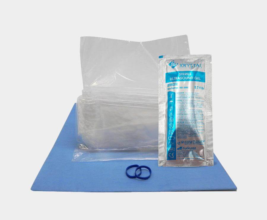
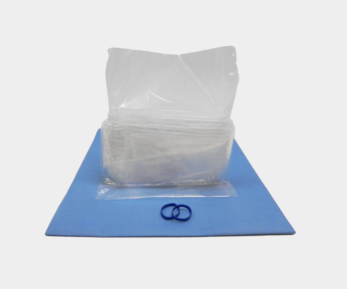
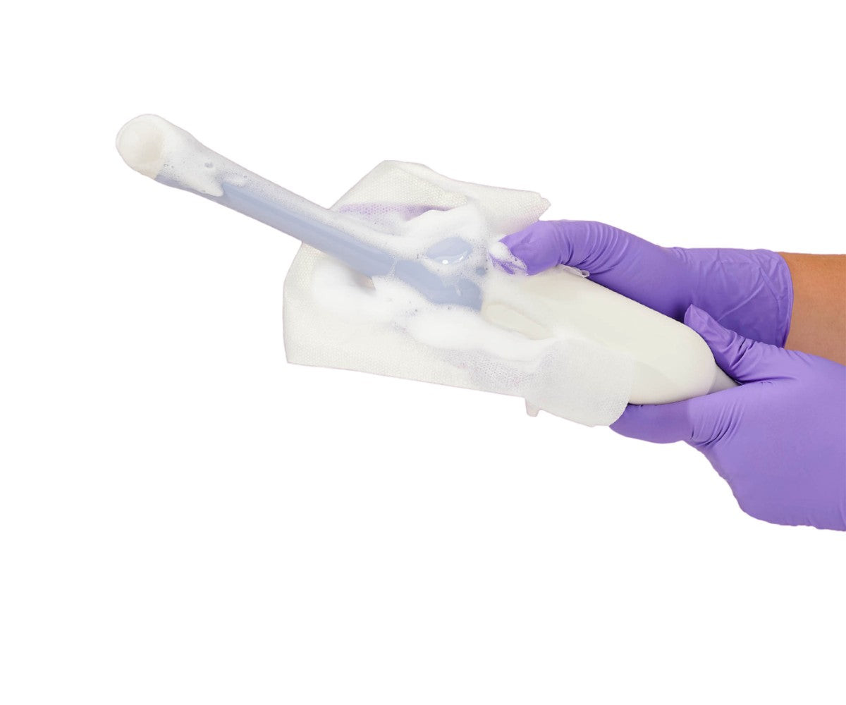
In certain cases, the sonographer or physician may opt to protect the transducer with a general purpose probe cover. This probe cover may be sterile or non-sterile. Its purpose is to prevent cross-contamination and is used to prevent the spread of healthcare-associated infections. For example, when performing breast biopsies, clinicians will protect the probe with a sterile cover as this is an invasive procedure and thus requires a higher level of precautionary measures.
The clinician will also apply ultrasound gel on the area being studied as ultrasound requires a liquid substance to operate. This may also be sterile or non-sterile. Many surface ultrasound exams will use a sterile gel; however, it is recommended that clinicians use sterile ultrasound gel for invasive procedures, such as biopsies.
Endocavity ultrasound scan
Endocavity ultrasound transducers have multiple applications: fetal, transrectal, transvaginal, and urological. For cancer-related concerns, physicians will employ endocavity ultrasound to perform biopsies in these areas. This allows for a more accurate retrieval of the sample.
Endocavity ultrasound transducers are protected with specially-designed endocavity probe covers. These probe covers lessen the risk of cross-contamination and increase patient safety. It is recommended that physicians use sterile probe covers for biopsies and other minimally invasive ultrasound-guided procedures. Non-sterile endocavity probe covers are also available. As in other ultrasound exams, the clinician will use ultrasound gel to perform the procedure.
Endoscopic ultrasound scan
Some cases may require an endoscopic ultrasound exam. These exams are minimally invasive imaging procedures. To perform the scan, the imaging professional will place the endoscopic transducer into the patient's mouth or rectum, instead of passing the transducer over the skin as is done with a surface ultrasound scan.
Endoscopic ultrasound transducers are typically protected with endoscopic probe covers to prevent cross-contamination. The clinician will use ultrasound gel in this procedure as well and may opt for sterility depending on the case.
 Figure showing an endoscopic ultrasound scan, courtesy of Educational Dimensions
Role of ultrasound in a cancer diagnosis
Although the clarity and details of the image produced by an ultrasound may not be the same as the MRI or CT scan, this imaging technique can still play an important role in the diagnosis of cancer.
The sound waves produced by the ultrasound echo differently from fluid-filled cysts and solid masses; therefore, an ultrasound exam can expose tumors that may be cancerous.
Further testing would be necessary to confirm cancer diagnosis, but ultrasound continues to be a viable option as an early cancer detection tool.


