Ensuring patient safety in imaging centers is not only an ethical obligation but also a key aspect of providing high-quality healthcare services. Patients entrust imaging centers with their well-being during various diagnostic and interventional procedures, necessitating a comprehensive approach to safety measures. In this article, we will explore useful tips and strategies for optimizing patient safety for imaging centers and radiology departments in general.
Ensuring Sterile Radiology Environments
The importance of maintaining a sterile environment within radiology departments cannot be overstated, as highlighted by a study revealing concerning levels of contamination in X-ray rooms. The study, published in 2014 in Acta Radiologica, found that a significant percentage of X-ray tubes (41.7%) and control panels and imaging plates (91.7%) were contaminated, posing potential risks to patients and staff.
According to the study, gram-negative organisms, notably Klebsiella pneumoniae, Pseudomonas aeruginosa, and Acinetobacter baumannii, were identified as common contaminants on X-ray machines, with the capability to persist on surfaces for extended periods.
In response to these findings, Professor Fatma Amer, PhD in her paper “Infection Prevention and Control in the Radiology Department” has recommended practices such as regular environmental cleaning of waiting areas, performing cleaning at least three times a day at fixed intervals, with terminal disinfection conducted using household bleach containing 5000 ppm available chlorine.
Additionally, spillages of blood or organic materials should be promptly addressed using a solution containing 10,000 ppm available chlorine to mitigate the risk of contamination and infection transmission among patients and staff.
It should be noted that spillages of blood and other bodily fluids not only pose a threat to patients but to the equipment itself also. For example, CT scanners used in interventional procedures are prone to the risk of being damaged over time if exposed to blood and bodily fluids that seep into the sensitive parts of the machine.
While radiology staff can and should wipe down machinery with approved cleaning and disinfectant solutions, that does not eliminate the risk of internal equipment damage. To address such concerns, it’s recommended that facilities drape their imaging equipment during interventional procedures in which bodily fluids may come into contact with the machine.
Proper draping of CT scanners and other imaging equipment enhances infection control and extends machinery lifetime, too. It mitigates the need for costly repairs and eventual replacement due to damage over the course of fluid exposure.

In addition to environmental measures, efficient sterilization and disinfection protocols for medical imaging equipment, depending on their usage, are imperative to uphold hygiene standards and prevent cross-contamination.
For example, radiographers and technicians should adhere to hand hygiene procedures outlined in the ISID's Guide to Infection Control in the Healthcare Setting and utilize hospital-approved alcohol wipes or chlorhexidine-based disinfectants to clean X-ray equipment and other instruments between examinations.
Surfaces directly in contact with patients should be covered with disposable sheets, such as MRI Non-Magnetic Poly Vinyl Chloride (PVC), which are replaced after each use to prevent transmission of pathogens. As noted prior, sterile equipment drapes are a recommended solution during interventional procedures to minimize the risk of pathogenic transmission and equipment damage. Non-sterile equipment drapes, or disposable sheets, may not be suited to handle blood and/or other bodily fluids.
Attention to detail is crucial, especially when decontaminating radiographic markers, with specific protocols recommended for ribbon markers due to their propensity for contamination.
Furthermore, the disinfection of MRI machines involves the use of chlorine-containing disinfectants or alternative agents, ensuring thorough coverage of all surfaces and adherence to prescribed disinfection times to effectively eliminate pathogens.
Ultimately, it is essential for imaging centers and radiology departments to collaborate with their facility’s infection prevention and control team. Increasing staff’s knowledge of infection control protocols through training and continuing education is a sure-fire way to ensure better adherence and increased patient safety.
Optimizing Radiation Protection
Radiation protection in medical imaging is a multifaceted endeavor aimed at safeguarding both patients and healthcare workers from the potential hazards of ionizing radiation.
Studies have demonstrated an increase in cancer risk among individuals, especially children and young patients, with previous exposure to CT scans. Additionally, measurable increases in radiation-induced DNA damage following various radiologic examinations have been observed, showing the importance of minimizing unnecessary radiation exposure wherever possible.

The European Commission's guidelines emphasize the importance of radiation protection education and training for medical imaging professionals, revealing a knowledge gap among healthcare professionals regarding radiation protection issues and radiation doses of commonly performed imaging procedures.
Radiographers, positioned as the final gatekeepers in the radiation protection chain, must undergo intensive education programs, attend radiation safety courses, and stay updated on new technologies to ensure patient safety.
Dr. Nicholas Frane, in his article “Radiation Safety and Protection,” mentioned that minimizing radiation exposure in medical settings involves implementing practical strategies such as increasing the distance between the x-ray beam and the patient or radiology staff. By positioning the image intensifier or x-ray plate as close to the patient as possible and the x-ray tube as far away as feasible while maintaining image resolution, radiation exposure can be significantly reduced.
This principle applies to clinicians as well, as scattered radiation encountered during fluoroscopy procedures follows the inverse square law, meaning exposure levels decrease with distance from the x-ray source. By doubling their distance from the source, staff can lower their exposure levels by a factor of four, highlighting the effectiveness of this simple yet impactful approach in minimizing occupational radiation exposure.
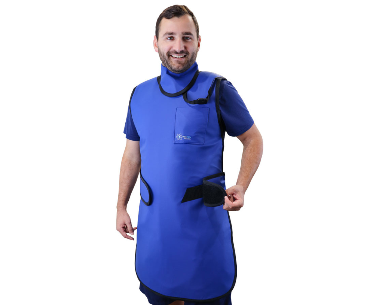
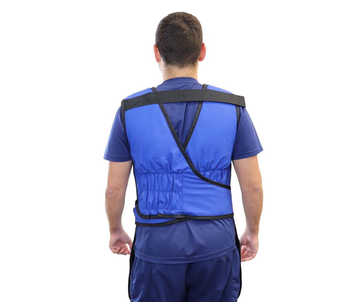
In addition to that, Dr. Frane also suggested using physical radiation shielding protecting both medical professionals and patients. Various forms of personal protective equipment (PPE), such as lead aprons and lead glasses, offer effective shielding against radiation exposure.

Radiation protection aprons, commonly available in different levels of thickness and various core materials, are essential for medical personnel working in fluoroscopy suites, operating rooms, and interventional settings. They should be accompanied by a thyroid shield for comprehensive protection. Furthermore, ensuring proper storage and testing of PPE is vital to maintain its effectiveness. Regular checks of lead garments every six months help ensure their integrity, while proper hanging of leaded aprons prevents cracking and ensures longevity.
Despite these measures, compliance rates for certain items, such as leaded eyeglasses, remain relatively low, highlighting areas for improvement in radiation safety practices. Studies have shown a clear relationship between occupational radiation doses and the development of cataracts, underscoring the significance of proper radiation protection equipment usage. Enhancing awareness and education regarding the importance of PPE in radiation protection is essential to promote better compliance among imaging center staff.
After all, implementing these measures ensures a safe environment not just for radiology staff but for patients as well, enhancing the quality of care provided. We encourage administrators and leaders in imaging centers and radiology departments to apply the tips and strategies discussed here to better optimize their practices.