Breast ultrasounds play an important role in the assessment of breast lumps and are used to determine whether they could be tumorous or fluid filled cysts. Doctors often request them as a precautionary measure, and in some cases, early detection could be a matter of life or death.
In a
recent study conducted in Sweden, researchers found that diagnostic imaging contributed to a 41% reduction in the mortality rate of breast cancer cases. Moreover, the authors of a
study published in February highlighted how ultrasound scans were able to identify 9 unique features linked to breast cancer.
Using high frequency sound waves, a breast ultrasound creates a black-and-white image of breast tissues and structures.
In this article, we will discuss the uses of breast ultrasound, what you can expect during the procedure, and how to best prepare for it.
Uses
As mentioned before, physicians commonly use ultrasound as an early diagnostic tool to evaluate lumps in the breast.
Ultrasound is unique from CT scans and X-rays in that it does not use ionizing radiation. Therefore, physicians will regularly recommend an ultrasound for patients who do not quality for radiation-based imaging techniques.
The following individuals should avoid radiation:
-
those who are pregnant or breastfeeding
-
those who are under the age of 25
-
those who have breast implants
Doctors may also use an ultrasound during a biopsy procedure. The ultrasound helps guide the biopsy needle to collect tissue from a lump for testing to determine whether it is malignant. The doctor may also employ a needle guide during the procedure to ensure precision and speed. This procedure is referred to as an ultrasound-guided biopsy.
 If the physician discovers a lump in the breast tissue during a routine physical exam or mammogram, then he or she may schedule a breast ultrasound to further investigate.
Breast ultrasounds may also be used for:
If the physician discovers a lump in the breast tissue during a routine physical exam or mammogram, then he or she may schedule a breast ultrasound to further investigate.
Breast ultrasounds may also be used for:
-
evaluating cases of mastitis, which is the inflammation of the mammary tissues
-
assessing unusual nipple discharge
-
monitoring breast implants
-
examining skin changes, such as discoloration
-
monitoring existing benign breast lumps
-
verifying the results of other imaging tests, such as an MRI scan or a mammogram
Preparation
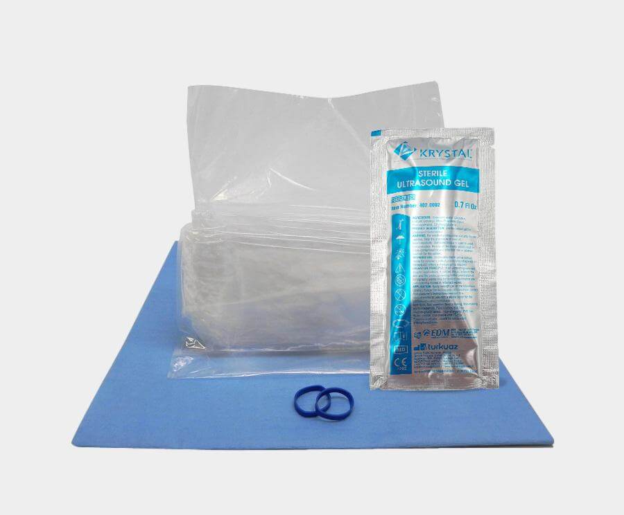
Patients should try not to apply any lotions or powders to their skin prior to the ultrasound exam. In addition, it is recommended that patients avoid wearing any metal, such as jewelry. All of these products may interfere with the results and decrease their accuracy.
The majority of breast ultrasounds are performed by sonographers in the radiology department of a hospital, although some might occur in a private or specialist clinic. Radiologists may sometimes perform them as well.
To avoid having to remove all clothing during the procedure, it is recommended that patients wear separate items of clothing on the top and bottom. For some individuals, wearing a button up or zippered shirt may also make the undressing process more comfortable.
What to expect
Breast ultrasounds are relatively short procedures. They typically last between 15 to 30 minutes and are also painless unless the lump is tender.
At the beginning of the process, the sonographer or radiologist will physically examine the breast and ask a series of questions about the lump:
-
when the patient noticed it
-
whether other symptoms are present
-
whether it has progressed
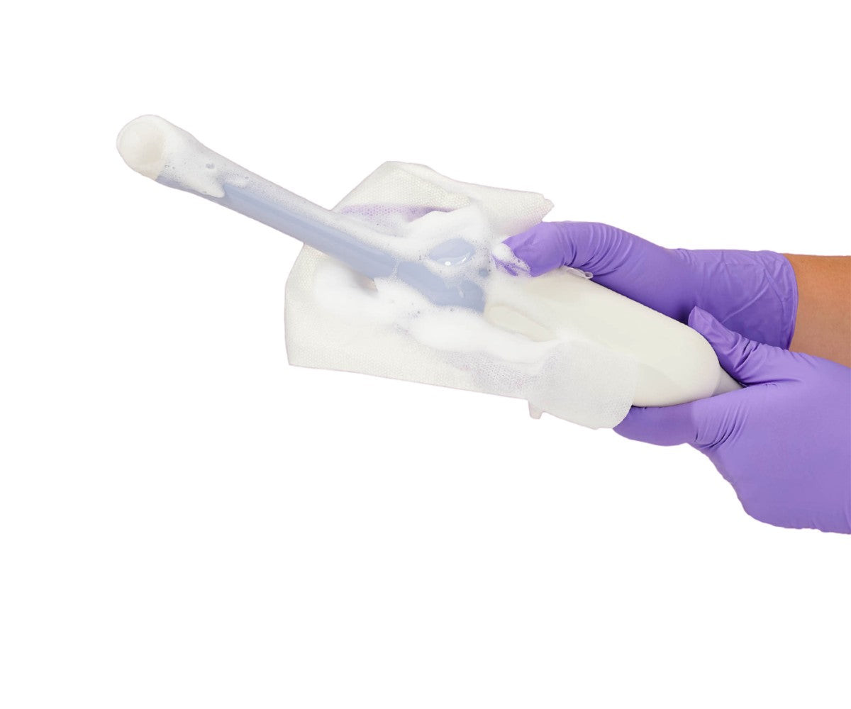
After the examination, the clinician will apply an ultrasound gel to the breast. By limiting air bubbles, the gel facilitates the ability of sound waves to move through tissues, thus providing a clearer image.
There are cases where the clinician will place a triangular pillow under the patient’s shoulder, causing the body to roll to one side. They may also raise the patient’s arm over their head. Both positions can make it easier for the sound waves to travel and for the tissue to receive them.
Once the sonographer has evenly spread the gel, they will pass a device called the transducer over the breast. This device sends sound waves through the breast and records their activity.
When a sound wave hits a tissue or structure during an ultrasound procedure, it bounces back. The ultrasound transducer then sends the following information to a computer: length of time it takes for the wave to bounce back, plus its volume, pitch, and frequency. The computer will then translate this information into an image referred to as a sonogram.
As the sonographer performs the ultrasound on the breast, he or she will examine all of its tissues and structures and take several images of different areas from various angles.
Moreover, it is common for them to create multiple still images of the lump and its surrounding areas. They may also take short moving videos.
After scanning the breast with the ultrasound transducer, the clinician will check the armpit for swollen or hard gland and nodes.
If a sonographer has any concerns about the accuracy of the results, then they may consult a radiologist for a second opinion. The radiologist may need to redo portions of the breast ultrasound to properly evaluate the area of concern.
Results
Although waiting for the results of a breast ultrasound can be stressful, it’s important to note that only few breast lumps pose a significant health risk.
For example, cysts typically don’t require any further treatment unless they are causing pain or are near blood vessels or breast ducts.
The results may be available the same day or a few days after the procedure, depending on the medical practice. In the majority of cases, the results from the breast ultrasound will go to a primary care physician who will call the patient to discuss them or explain them during an office visit.
There are other factors which can influence how quickly the results are available:
-
the potential risk of symptoms and the need for prompt treatment
-
whether the radiologist needs more information to interpret the results
-
whether the doctor needs to compare the results to previous ultrasounds or the results of other imaging tests, such as a CT scan or X-ray
-
whether the symptoms could be the result of an underlying condition
-
whether the facility is delivering the results by email, fax, phone, or post
Fluid and solid tissue appear differently on a sonogram. If the breast ultrasound confirms that the lump is a fluid filled cyst, then the physician may discuss the results over the phone.
If the ultrasound finds a solid lump, the imaging results alone will not be enough to confirm or rule out cancer or other conditions. Usually, the doctor will then request an MRI and biopsy to evaluate abnormal breast ultrasound results.
In the case of inconclusive or abnormal results, it is helpful to keep in mind that cancer is not the only health condition that causes breast lumps.
Other common causes of noncancerous breast lumps include:
-
Cysts – a fluid-filled sac in the breast
-
Fibrocystic breasts – noncancerous changes that give a breast a lumpy or ropelike texture
-
Fibroadenoma – a noncancerous breast tumor that most often occurs in young women
-
Fatty lumps comprising bruised, dead, or injured mammary fat cells
-
Hyperactive breast glands – occurs when there’s an increase in the number of cells lining the ducts or lobules of the breasts
-
Intraductal papilloma – a wart-like lump that develops in one or more of the milk ducts in the breast
-
Hormone conditions, changes, or therapies
-
Certain medications – such as birth control pills
-
Mastitis or breast infection

 Ultrasound gel being applied to a transducer