According to a recent study published on February 24th in Ultrasound in Medicine and Biology, investigators have found nine unique patient and ultrasound features that may enable breast imagers to distinguish benign from malignant complex cystic or solid breast lesions.
Ultrasound is used for a wide variety of applications in women's health, such as ultrasound-guided breast biopsies and transvaginal ultrasound (TVUS) exams.
The research revealed that lesion type, orientation, size, and vascularity were some of the features found via handheld ultrasound scans and all were linked to breast cancer. Investigators reported that the combination of all nine features resulted in a sensitivity of 77% and a specificity of 82%.
"In addition to identifying whether a lesion is cystic
or solid,
The study defined complex cystic lesions as those that are round or oval in shape with circumscribed margins but that also feature debris with low-level echoes. Although they are commonplace in middle-aged women and often benign, these lesions still carry a nontrivial risk of malignancy. Therefore, the research sought to answer the question of whether features on handheld ultrasound might be better equipped to differentiate complex cystic lesions which required biopsy and others which were likely benign.
In an effort to find the answer, researchers used data from 453 patients with 472 complex cystic and solid breast lesions who visited a university cancer center between 2000 and 2018. The lesions were examined using handheld ultrasound with a high-frequency linear array transducer by radiologists with at least three years of breast imaging experience.
The researchers subdivided the lesions into four categories:
- Type I: Thick wall and/or septations, > 0.5 mm
- Type II: One or more mural or papillary nodules
- Type III: Mixed lesion that is more than 50% cystic
- Type IV: Mixed lesion that is more than 50% solid
The study included a variety of lesion types. About 13% of the lesions were classified as type I, 10% as type II, 21% as type III, and 55% as type IV.
The research found that following biopsy a little more than half of the lesions were confirmed as benign. Meanwhile, the remaining 45% were deemed malignant. It should be noted that the results varied on the basis of lesion type.
The positive predictive value was found to be much higher for type III or type IV lesions than for type I or type II. Investigators also reported a number of other factors independently associated with malignancy:
- Lesion diameter of more than 18 mm
- Lesion with irregular shape
- Nonparallel orientation
- Uncircumscribed margin
- Calcification
- Vascularity in the tumor
- Abnormal axillary lymph nodes
- Patient age of 52 and older
The study noted how a lesion type of III or IV and a lesion diameter of more than 18 mm had the highest level of sensitivity (87%) and negative predictive value (82%) of the factors associated with malignancy. Furthermore, abnormal findings in axillary lymph nodes had the highest level of specificity (94%) and positive predictive value (83%).
Although type I and type II lesions could not be ruled out as malignant since they met the 2% malignancy threshold to qualify for a BI-RADS 4, the researchers noted that they were able to better categorize them.
“Type I, II, and IV lesions can be assessed as BI-RADS 4B, while type III lesions can directly go into BI-RADS 4C, which allows a more accurate subclassification of complex cystic and solid breast lesions in clinical practice,” the researchers noted.
It is important to note that the study may have selection bias as the researchers excluded complicated cystic lesions without solid components and elected to solely include patients who had already undergone a handheld ultrasound.
Regardless of the potential for selection bias, the investigators believe the findings reaffirm the viability of handheld ultrasound as a formidable, secondary screening tool.
They surmised the study by writing, "Therefore, we may safely conclude that the
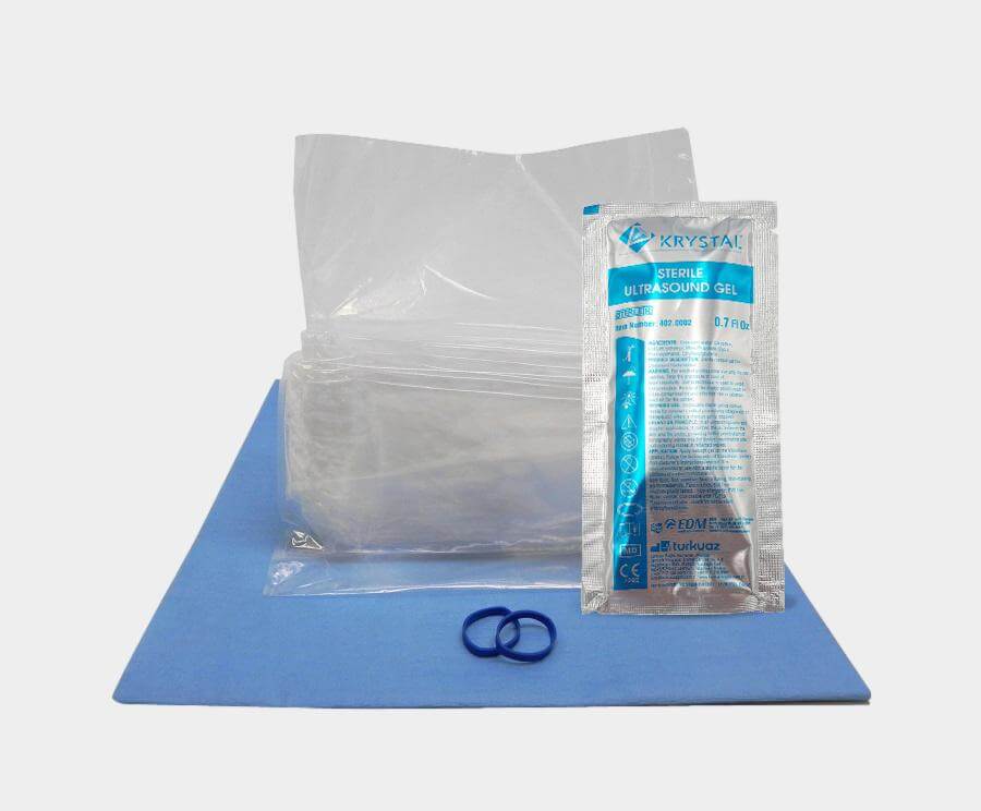
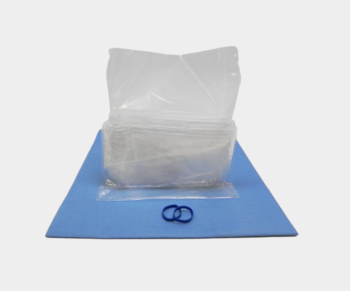
EDM offers supplies and equipment for ultrasound-guided procedures, such as biopsies. Our sterile ultrasound probe cover protects patients and medical staff from the risk of healthcare-acquired infections. We also offer sterile ultrasound gels that provide a highly-conductive solution while improving patient safety, such as the EDM sterile ultrasound gel.