Breast cancer impacts the lives of millions of women each year in the United States and continues to be a foremost topic in women’s health. Recent estimates state that 1 in 8 women (about 12%) in the US will develop invasive breast cancer over the course of her lifetime. This year alone, it is estimated that there will be 276,480 new cases of invasive breast cancer, and 48,530 new cases of non-invasive (in situ) breast cancer.
As a preventative measure, physicians commonly perform a variety of exams for early detection. These can be performed via mammography, physical examination, or other imaging studies. The lumps or abnormalities typically associated with breast cancer are most often detected during one of these exams.
Researchers have noted how ultrasound scans provide a quick, efficient, and cheap way to perform breast examinations. Although imaging modalities face hurdles in certain groups of patients, such as those with dense breasts or tissue abnormalities, Dr. Veronica Girardi from the Istituti Ospedalieri Bresciani in Brescia, Italy, advocates the use of ultrasound because when compared to mammography and MRI, it proves to be a well-tolerated, cheap, and safe option, she says. Moreover, researchers recently found that handheld ultrasound scans were able to identify nine distinct features linked to breast cancer.
But to determine whether the growth or abnormality is benign or cancerous, imaging tests alone may not be sufficient.
This is where breast biopsies come into play. When medical imaging proves insufficient to give a proper diagnosis, physicians will order a biopsy to be performed on the area in question. During the biopsy, some cells will be removed from the area in the breast and then be examined under a microscope for diagnosis.
Breast biopsies can be performed surgically; however, it is more common for the procedure to be performed by a radiologist in a less invasive fashion by using a hollow needle and image guidance. It should be noted that an image-guided needle biopsy is not meant to remove the entire lesion from the breast, but rather intended to retrieve a small sample of the lump or abnormality for further analysis.
Image-guided breast biopsies are performed by retrieving samples of an abnormality with the assistance of a medical imaging modality, such as MRI, ultrasound, or mammography. Radiologists may commonly turn to ultrasound-guided breast biopsies due to ease of use and lack of radiation, which is one of the key differences between ultrasound and other imaging modalities such as MRI and CT scans.
Common Uses of Ultrasound-Guided Breast Biopsies
Ultrasound-guided breast biopsies are often performed when a breast ultrasound shows an abnormality.
Breast biopsies are known to be very reliable exams. As the University of Tennessee Medical Center reports, "Breast biopsy procedures rarely miss a lesion, or underestimate the extent of disease present." Therefore, physicians often resort to breast biopsies when other forms of examination alone cannot determine the presence of cancer.
These abnormalities include but are not limited to:
- a suspicious solid mass
- a distortion in the structure of the breast tissue
- an area of abnormal tissue change
In cases where the mass can be felt, the physician may still opt to use imaging guidance.

Ultrasound guidance is primarily used in four types of biopsy procedures:
- Fine needle aspiration (FNA): this procedure uses a very small needle to retrieve fluid or cells from the suspicious area.
- Core needle (CN): this procedure involves a large, hollow needle to remove one sample of breast tissue per insertion.
- Vacuum-assisted device (VAD): the physician will use a vacuum-powered instrument to collect multiple tissue samples during one needle insertion.
- Wire localization: this procedure involves placing a guide wire in the suspicious area to assist the surgeon in locating the lesion for a surgical biopsy.
How can patients prepare for a breast biopsy?
It is recommended that patients wear comfortable, loose-fitting clothes for the procedure. Patients may need to remove all clothing and jewelry in the area being examined. Additionally, patients may be asked to wear a gown.
Before the biopsy, patients should inform their physician of any and all medications they are taking, including herbal supplements. It is also recommended that they inform the physician of any allergies, particularly if the patient is allergic to anesthesia. The doctor may advise the patient to stop taking aspirin, certain herbal supplements, or blood thinners three to five days prior to the procedure to reduce the risk of bleeding.
Lastly, practitioners should remind patients to inform their physician of any recent illnesses or other medical conditions. Patients may be sedated during the biopsy; therefore, medical staff should recommend that they bring along a relative or friend who can drive them home afterwards.
Equipment used during an ultrasound-guided breast biopsy
During the procedure, a medical imaging practitioner, typically a radiologist, will utilize an ultrasound transducer (also known as a probe) and a needle to perform the biopsy.
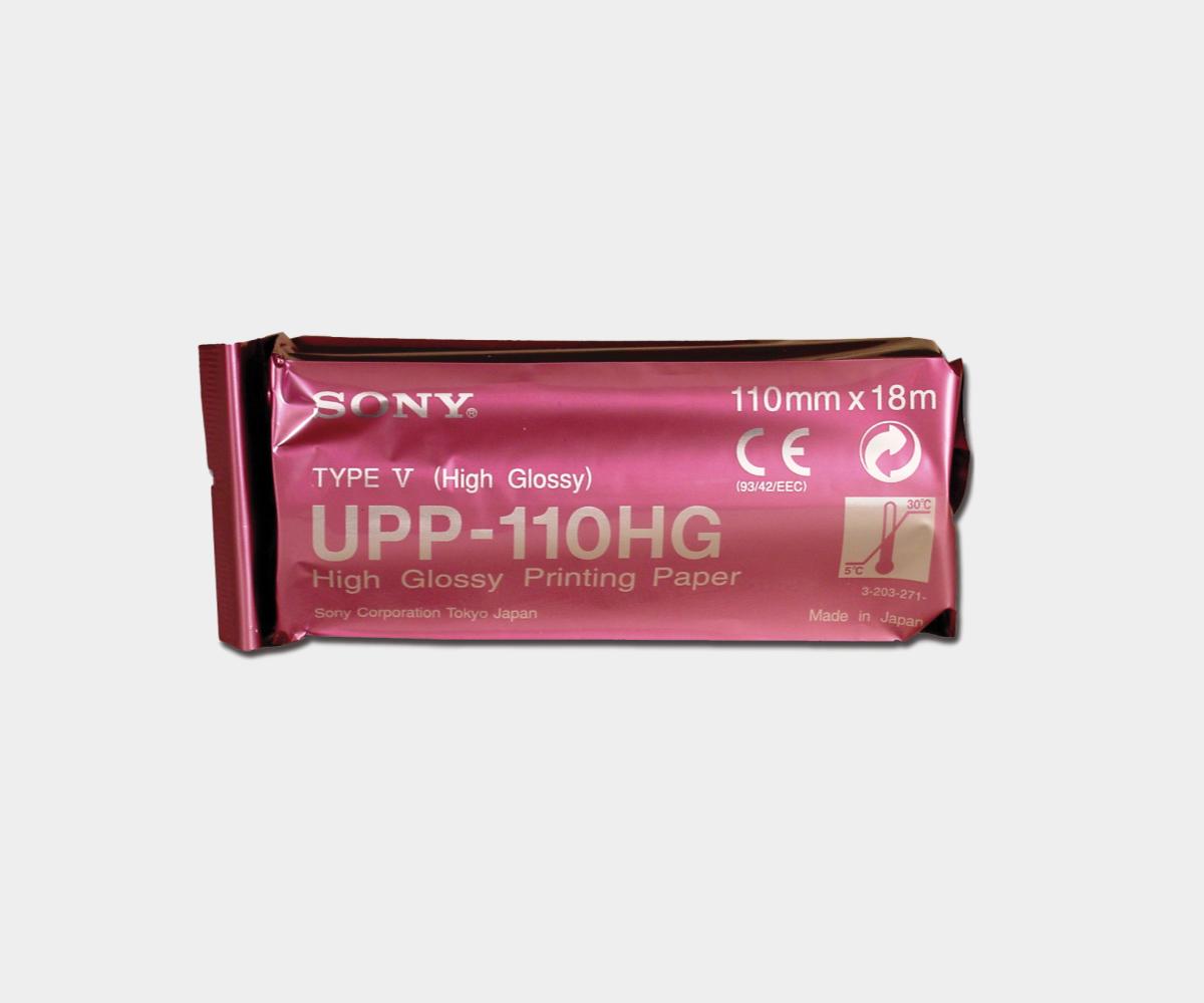
After applying a sterile ultrasound gel to the examination area, the radiologist will press the transducer onto the skin and begin to scan the area. The probe sends sound waves and records the echoing waves. The sensitive receiver in the transducer records minute changes in the sound’s pitch and direction. These waves are then measured by the ultrasound machine and displayed by a computer. Thus, the waves are converted into a real-time image which the radiologist can follow as he or she performs the procedure.

To this end, the ultrasound provides the clinician with a visualization of the location of the breast mass as well as any distortion or abnormal tissue change. Once the radiologist identifies the location of the mass, he or she will insert the biopsy needle through the skin and towards the targeted finding. When the needle reaches its target, the radiologist will then remove various tissue samples for diagnosis.

In the case of a surgical biopsy, the ultrasound may be used to guide a wire into the target, so that the surgeon can locate the area for excision with greater ease when the procedure is performed. It should be noted that the wire is placed in the target area hours before the surgery takes place. The image-guided procedure is helpful for physicians as it allows them to view the biopsy needle or wire as it advances to the location of the lesion or abnormality in real-time, thereby increasing procedural efficiency, precision, and ultimately, patient safety and comfort.

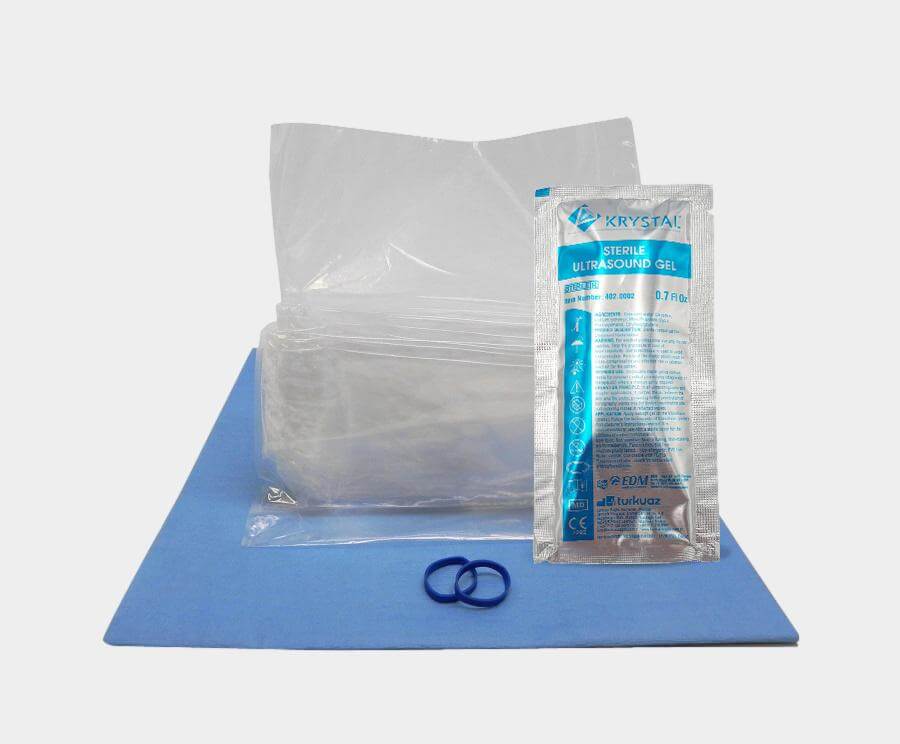
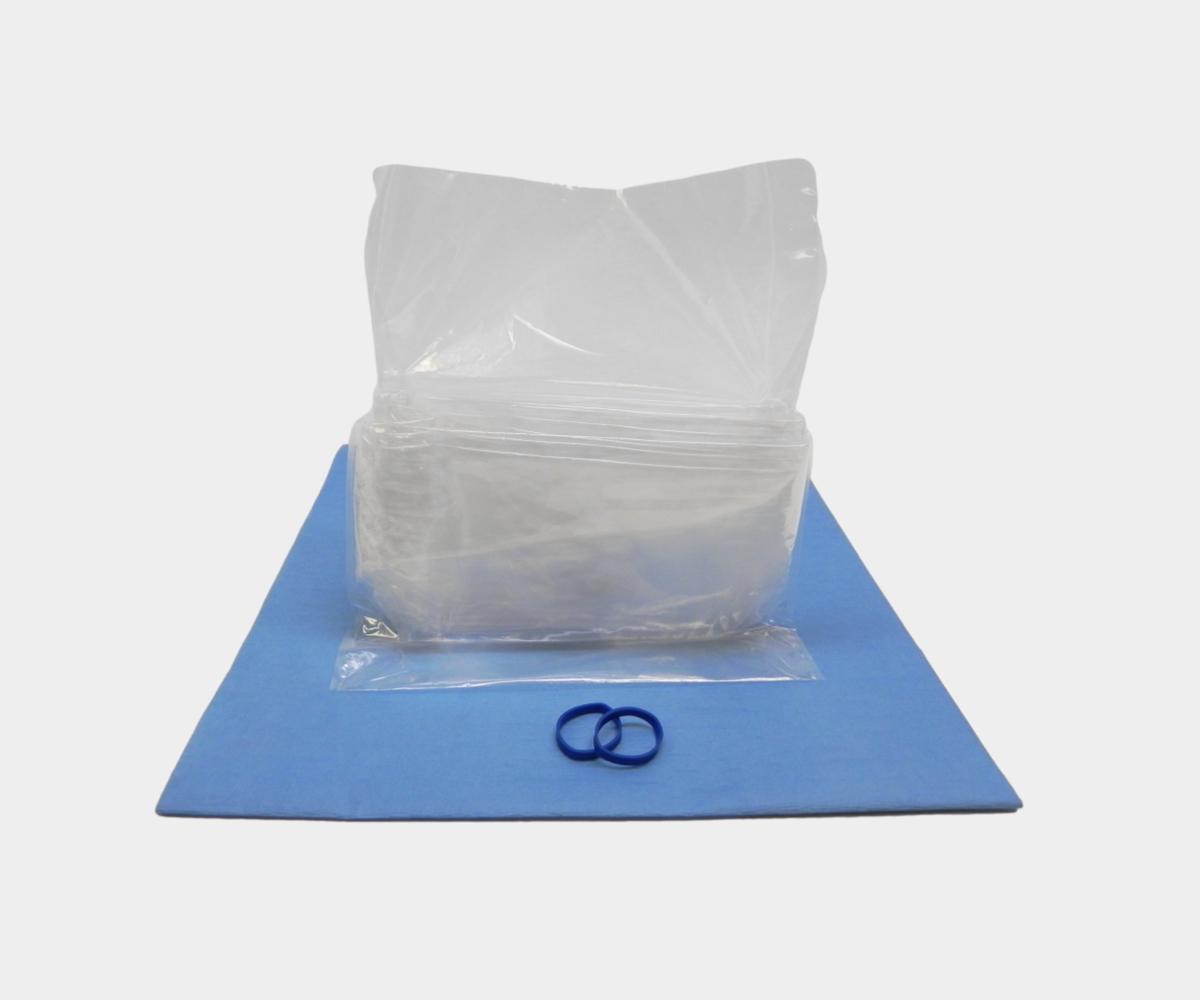
To reduce the risk of cross contamination, physicians may utilize a sterile surface probe cover to create a barrier of protection between the ultrasound transducer and patient. These covers preserve the sterile field and also protect the transducer by not allowing it to be contaminated.
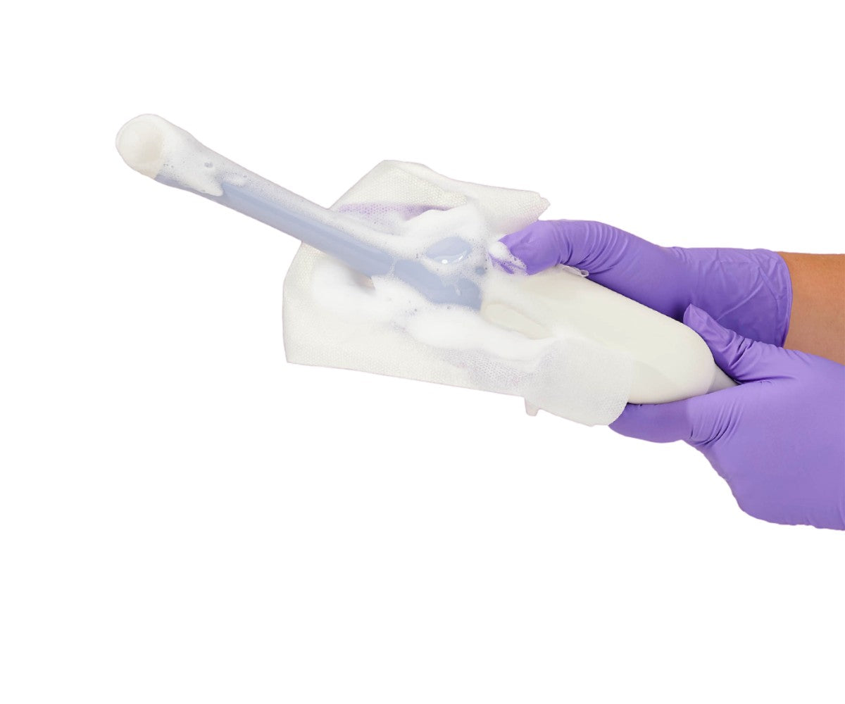
In addition, medical staff should clean and disinfect the probe with hospital-grade products, such as disinfectant sprays and wipes, before and after each patient use to reduce the transmission of healthcare-associated infections.
How is an ultrasound-guided breast biopsy performed?
Unlike surgical breast biopsies, ultrasound-guided breast biopsies are often times minimally invasive procedures which are usually performed by a specially trained radiologist. In most cases, these biopsies are done as an outpatient procedure.
The patient will be positioned lying face up on the examination table, or turned slightly to one side. Afterwards, the clinician will inject a local anesthetic into the skin to numb the breast. The radiologist will then proceed to apply a sterile ultrasound gel and use the transducer to locate the lesion. Once the lesion has been located via ultrasound, the radiologist will make a small nick in the skin at the site where the biopsy needle will be inserted. While monitoring the examination area with the ultrasound probe, the clinician will insert the needle and advance it towards the target site.
When the needle has reached the mass, the tissue samples will be removed via one of three methods:
- Fine needle aspiration: a fine gauge needle and a syringe are used to retrieve fluid or clusters of cells. The Mayo Clinic calls FNA the "simplest type of breast biopsy" due to its thin needle and rather quick procedure time. FNA is an efficient way to distinguish between a fluid-filled cyst and a solid mass, and can help avoid a more invasive procedure.
- Core needle biopsy: the clinician activates the needle’s automated mechanism, which moves the needle forward and fills the needle trough (also known as a shallow receptacle) with “cores” of breast tissue. The outer sheath of the needle moves forward instantly to cut the tissue and store it in the trough. This is repeated about three to six times depending on the biopsy.
- Vacuum-assisted device (VAD): The tissue is pulled from the target area utilizing vacuum pressure via the needle. This tissue is then deposited in a sampling chamber. This needle does not require withdrawing and reinsertion as it can rotate positions and collect multiple samples. Eight to ten samples of tissue are often collected from the target area.

Following the extraction of the samples, the biopsy needle is removed. The radiologist will apply pressure to stop any bleeding that may occur following the removal of the needle and then proceed to cover the skin with a dressing. Sutures are not required.
The results will then be sent to a lab for histological analysis. The specimen will be examined by a pathologist who will then make a final diagnosis.