Ultrasound is generally considered one of the safest imaging modalities available today. For example, it does not expose patients to harmful radiation in the way computed tomography and x-rays do. It has also become a highly mobile modality. The surge of point-of-care ultrasound has given clinicians an increased ability to take medical imaging outside of the confines of the traditional imaging suite. Clinicians can now perform scans in areas where it once would have been nearly impossible to do so, including make-shift hospitals and helicopters.
The modality has grown in use across a wide range of clinical areas as well. While many in the general public may still associate ultrasound with a sonogram, clinicians across a multitude of fields have found its benefits. This includes those in radiology, obstetrics, cardiology, urology, and more. In addition to diagnostic applications, ultrasound is increasingly used to guide minimally invasive procedures. For example, endocavity ultrasound probes are often paired with endocavity needle guides to assist with biopsies, aspirations, and other interventional procedures. These needle guides attach directly to the probe, providing a fixed pathway that helps clinicians visualize and accurately advance a needle to a target structure in real time, improving precision and procedural safety.
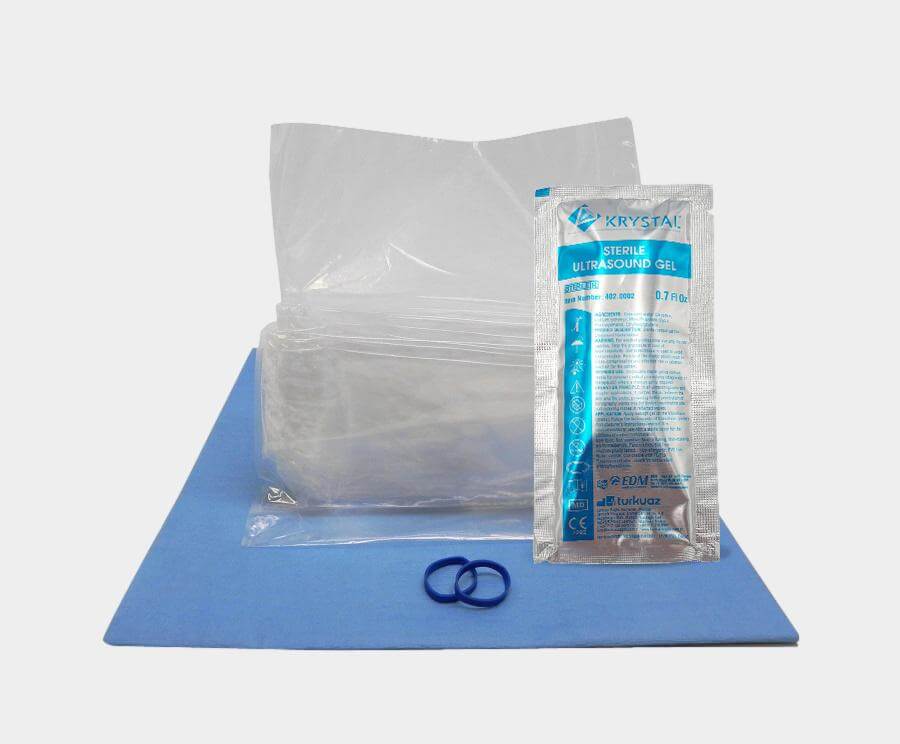
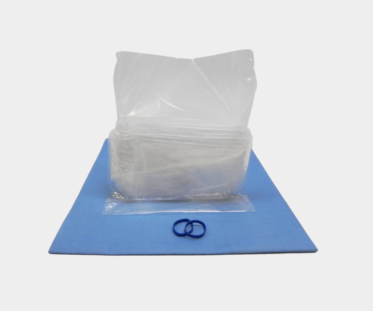
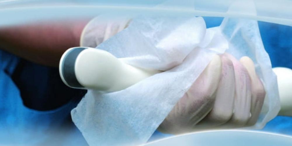
However, while such advances offer both clinicians and patients a series of benefits, ultrasound does not come without its risks. Endocavity ultrasound involves a transducer that comes into contact with the anal cavity, the vagina, or the oral cavity. When needle guides are used in these procedures, they too enter the sterile field and may come into close proximity with mucous membranes or sterile tissues. These areas are composed of mucous membranes and therefore the introduction of a non-sterile instrument—including an endocavity ultrasound probe or an improperly reprocessed needle guide—has important infection control implications. It is essential for patient safety that practitioners understand how proper cleaning, high-level disinfection or sterilization (as appropriate), and strict compliance with ultrasound hygiene protocols for both probes and accessories contribute to the prevention of healthcare-associated infections.

In a survey carried out by the European Society of Radiology (ESR), researchers found that 29% of respondents - who were ESR members and mostly radiologists working in large hospitals - did not disinfect the ultrasound probe after each patient. Such non-compliance is especially dangerous when utilizing an endocavity probe which enters sterile areas of the body. While ultrasound’s popularity has been fueled by its reputation for safety, this does not signify that practitioners should lower their guard when considering the value of reprocessing the probe.

Disinfect the entire endocavity ultrasound device
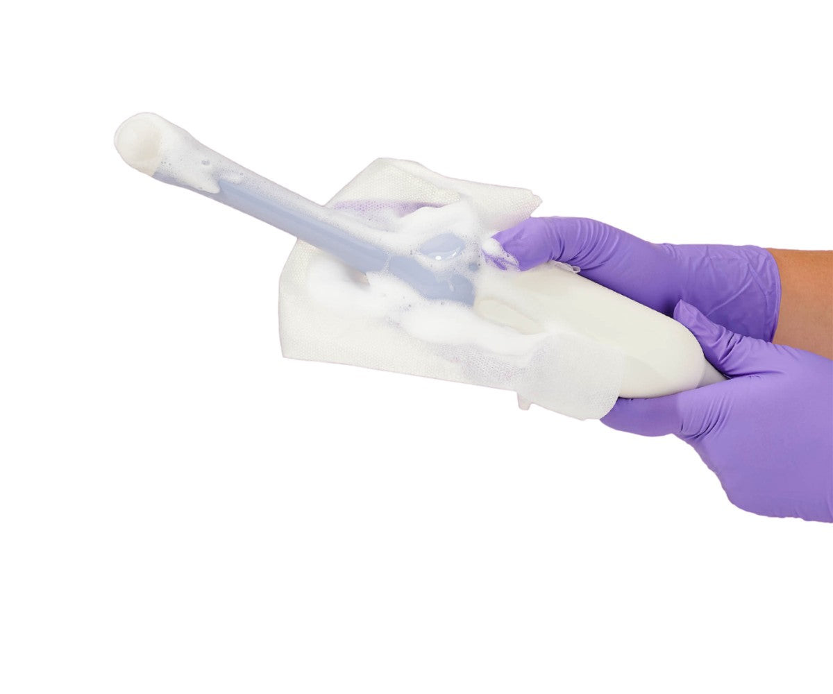
While some practitioners may be sure to disinfect the probe, it is sometimes easy to look over the other parts of the machine. However, ultrasound users must note that every part of the device should be viewed as a vector of infection. Transducer handles are one area that are commonly left out of the disinfection process. In an article published in the Australasian Journal of Ultrasound in Medicine, researchers noted this, “current ultrasound probe reprocessing guidelines do not specify inclusion of the transducer handle in the HLD process.” Failing to disinfect the handle is a mistake which clinicians must avoid.
The importance of this is evidenced in a study which found that 80.5% of vaginal ultrasound device handles were contaminated with bacteria right after probe immersion in glutaraldehyde during the HLD process. Researchers conducting the study reported that S. aureus was isolated in 15.4% of these samples. One isolate was also found to be methicillin‐resistant Staphylococcus aureus (MRSA).
“Causative agents of urinary tract infections were also isolated from the handle including Staphylococcus haemolyticus, Staphylococcus saprophyticus and Enterococcus faecium,” the study states.
The good news is that disinfection can assist in combatting the bacteria found on the handle. The researchers reported that handle contamination was reduced to “background levels” once disinfection of the handle was performed simultaneously with the probe body.
Therefore, clinicians must ensure that both the probe body and handle undergo HLD during endocavity ultrasound procedures. The handle must also be viewed as a vector of infection as well as a potential source of cross-infection. Depending on the procedure, some patients may come into direct contact with the handle or indirect contact via the practitioner or probe cover. Failing to disinfect the handle can thus cause the sterile field to be compromised and can promote the propagation of healthcare-associated infections.
Not all high-level disinfectants kill HPV
Clinicians must also be aware that not all high-level disinfectants offer the same degree of infection prevention. The human papillomavirus (HPV) is clinically significant to ultrasound usage as there is a notable overlap between HPV-related cancers and the body sites where endocavity procedures occur.
The results of a study conducted in 2014 revealed that glutaraldehyde and ortho‐phthalaldehyde (OPA) were rendered completely ineffective against native HPV16 virions. Even with extended contact times of up to 24 hours, both high-level disinfectants were unable to combat the contaminant. These alarming results should be kept in mind when clinicians choose which high-level disinfectant to use on their endocavity ultrasound probe. Many facilities continue to use these agents to disinfect endocavity probes and their usage should be reconsidered.
Other studies have also found residual HPV virus on transvaginal ultrasound probes before and after low-level disinfection performed with quarternary ammonium wipes. This highly stable virus can survive in a dry room for several days. As a result, clinicians must be cautious to not use an ineffective disinfection method as the risk of transmitting cancer-causing HPV to the following patient is only amplified through failed disinfection.
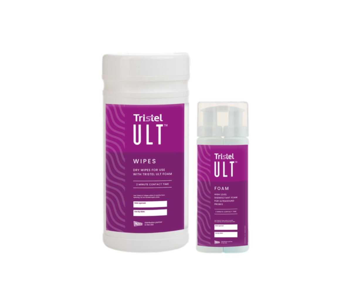
Ultrasound practitioners are not without options. A study found that a hydrogen peroxide-based disinfection system designed to perform HLD on ultrasound transducers was able to successfully and completely inactivate native HPV16 and HPV18 virions. Thus, ultrasound users should seek options such as this as a way of mitigating the risk of HPV transmission during their endocavity procedures, especially transvaginal ones.