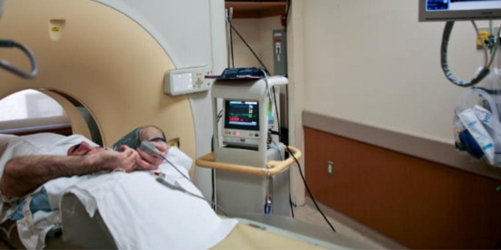
Radiation protection remains a top-of-mind concern within interventional radiology. When conducting CT scans, it is important that radiologists take into account the components of CT systems which can be used to lower the amount of radiation exposure. In particular, those conducting cardiac CT angiographies can utilize these components to reduce x-ray dose.
In an effort to combat exposure to radiation, several cardiology societies jointly published a consensus document in 2018 which outlined a series of best practices for x-ray dose reduction during coronary computed tomography angiography (CCTA) exams. The participating organizations include the American College of Cardiology (ACC), the Heart Rhythm Society (HRS), the North American Society for Cardiovascular Imaging (NASCI), the Society for Cardiovascular Angiography and Interventions (SCAI), the Society of Nuclear Medicine and Molecular Imaging (SNMMI), and the Society of Cardiovascular Computed Tomography (SCCT).
Below is a checklist which summarizes these best practices. By taking these steps, medical imaging professionals conducting these exams will be able to better reduce patient radiation dosage when conducting cardiovascular CT.
-
Patient Case Selection
Radiologists should consider the patient’s age, co-morbidities, and natural life expectancy. In addition, the appropriateness and utility of non-radiation-based imaging techniques should also be taken into account.
-
Equipment Calibration
Clinicians should use acquisition detector doses as low as compatible with the image quality.
-
Procedural Planning
Depending on the study goals, it is recommended that radiologists select the lowest-dose acquisition protocol. When appropriate, choose retrospective gating. In the case that retrospective gating is selected, utilize ECG-gated variable tube output. Moreover, use the lowest X-ray tube voltage and current compatible with diagnostic quality image acquisition. It is advised that clinicians use topogram-based tube current modulation. Lastly, use the largest scan pitch that works with diagnostic quality image acquisition.
-
Study Conduct
During the exam, radiologists should seek to minimize the patient’s heart rate. It is recommended that clinicians confine scanned body area to the area relevant to the diagnostic purpose of the study.
Factors Affecting Patient CT Dose

The jointly issued document sheds light on a variety of factors which affect patient CT dose. The radiation dose experienced by a patient is a result of a combination of the patient’s physical characteristics as well as scanner protocol selection. For example, depending on patient size and body mass index (BMI), patient X-ray exposure may need to increase to ensure diagnostic-quality images are acquired.
Patients who are larger and have a greater BMI will likely experience a higher dose than those who are smaller and have a lower BMI. However, according to the aforementioned guidelines, patient size is not a variable which determines exam appropriateness so long as the patient’s size does not prevent the acquisition of diagnostic-quality images. In addition, depending on the specific acquisition parameters, the increased X-ray exposure does not have to increase dose to radiosensitive tissues.
Furthermore, CT systems can use either a constant X-ray tube output or an ECG-gated variable output. This depends on the selected acquisition protocol. It is important to note that the radiologist, or operator, needs to select the acquisition protocol in accordance with patient characteristics and the purpose of the study. The objective of the operator should be to deliver sufficient X-ray exposure to allow for an acceptable degree of noise in the images.
Additionally, the document authors remind readers that the CT system operator has the responsibility of selecting a scanning protocol which optimizes the exam’s diagnostic yield while also minimizing CT dose.
Radiologists should take the following factors into consideration during this process:
Scan Length
The scan length is defined as the distance imaged along the cranio-caudal axis. It should be kept to a minimum to include solely the anatomy of interest. Operators should be careful to not expose structures which are not relevant to the study’s purpose. In addition, ensure that the diaphragm position seen on the topogram is the same as during scanning.
X-ray Beam Intensity
This is determined by both the X-ray tube potential (in units of kV) and the X-ray tube current (in units of mA). Today’s CT scanners will modulate the tube current dynamically throughout the CT acquisition to reduce radiation exposure.
Tube Potential
The adjustment of X-ray tube voltage has been found to be the most important factor in controlling X-ray dose. The jointly issued document stated that increasing tube voltage also increases the X-ray beam’s mean photon energy level, and thus, it increases radiation dose almost proportionally to the square of the voltage. This signifies that a decrease of tube voltage from 120 to 100 kilovolts (kV) reduces the radiation dosage by nearly 40 percent with a constant tube current. Note that the tube voltage in the majority of CT scanners can be adjusted between 70-140 kV and this voltage is selected by the machine operator in accordance with the patient’s weight or BMI.
Tube Current
This is defined as the number of electrons accelerated across the tube per unit of time and is proportional to the number of X-ray photons produced per unit time, according to the document authors. It should be noted that the radiation dose is linearly proportional to the tube current. Additionally, the image noise is inversely proportional to the square root of the tube current. Therefore, if operators decrease tube current at a given tube potential then they can decrease the dose at the expense of increased image noise. Depending on patient size, the tube current may be modified by the radiologist. The majority of modern CT systems allow for tube current modulation on the basis of the thickness of the patient’s body estimated from the topogram. This modulation may be applied longitudinally or circumferentially. Applying this approach can reduce radiation exposure of thoracic CT examination by 20 percent without increasing image noise.
Rotation Time
This is defined as how much time is needed for the gantry to perform one rotation. Additionally, radiation exposure increases linearly with rotation time.
Scan Acquisition Mode
The authors of the document explained how scan acquisition mode is a major determinant of radiation dose. Depending on the acquisition mode selected, patient exposure can be substantially different while producing similar images. The three main CT scan modes are axial or “conventional” scanning, fixed table or single-station scanning, and helical scanning.
Cardiac Motion Compensation
This is rarely applied outside of direct cardiac and aortic root imaging. As a result, the majority of cardiovascular imaging does not use ECG gating or triggering. However, when radiologists are imaging the heart or aortic root, cardiac root compensation is essential to avoid the presence of motion-related artifacts that reduce diagnostic image quality. CT operators can choose from two cardiac compensation methods depending on the scan mode. These methods are prospective ECG triggering and retrospective gating.
ECG-triggered Tube Current Modulation
This is utilized by radiologists to reduce radiation dosage during systole when there is the greatest cardiac motion. According to the document authors, it is capable of reducing radiation exposure to a great degree. During this technique, the tube current is only at nominal value during the portion of the cardiac cycle most likely to be used for reconstruction. The tube current is then reduced to decrease radiation output during the remainder of the cardiac cycle. Document authors stated that refinements of this technique have permitted a reduction of the window during which tube current is nominal as well as a decrease of tube current during the undesired portions of the cardiac cycle by 20 percent. However, CT operators should note that one disadvantage when employing ECG-triggered tube current modulation is that images reconstructed from projection data acquired with low tube current may result in images that are too noisy to be diagnostic for coronary anatomy.
Image Reconstruction
In the past, filtered back projection has historically been employed for the reconstruction of CT images. However, the development of greater computing power has given way to an alternative statistical method of iterative reconstruction practical for CT. Utilizing this method allows for the prediction of projection data based upon an initial assumption in regard to attenuation in each voxel and compares this data to measured projection data. Then the voxel attenuation values are modified iteratively until an acceptable error level between the measured data and the predicated data is acquired. The result is reconstructed images with lower noise values compared with those obtained with filtered back projection. Furthermore, using this method reduces tube voltage and/or tube current to acquire images with a lower radiation dose and comparable noise.
Image Post-Processing Filters
CT operators may also consider applying image post-processing filters to reduce radiation dose. They can be applied to images to reduce their noise while still maintaining image contrast and edges. The document authors noted how the feasibility of using these filters for exposure reduction has been recently demonstrated.
In addition to these scanning techniques, it's important to note that CT operators and interventional radiologists have a wide array of radiation protection equipment at their disposal. Learn more about how radiation protection aprons and accessories can keep clinicians and patients safe here.